A 115‑million‑year‑old dinosaur bone with a hint of blood
Did any trace of a dinosaur’s blood survive deep time, and if so, how would we know for sure? That’s the blunt question behind a new study that hunts for a chemical calling card of red blood cells in a 115‑million‑year‑old leg bone.
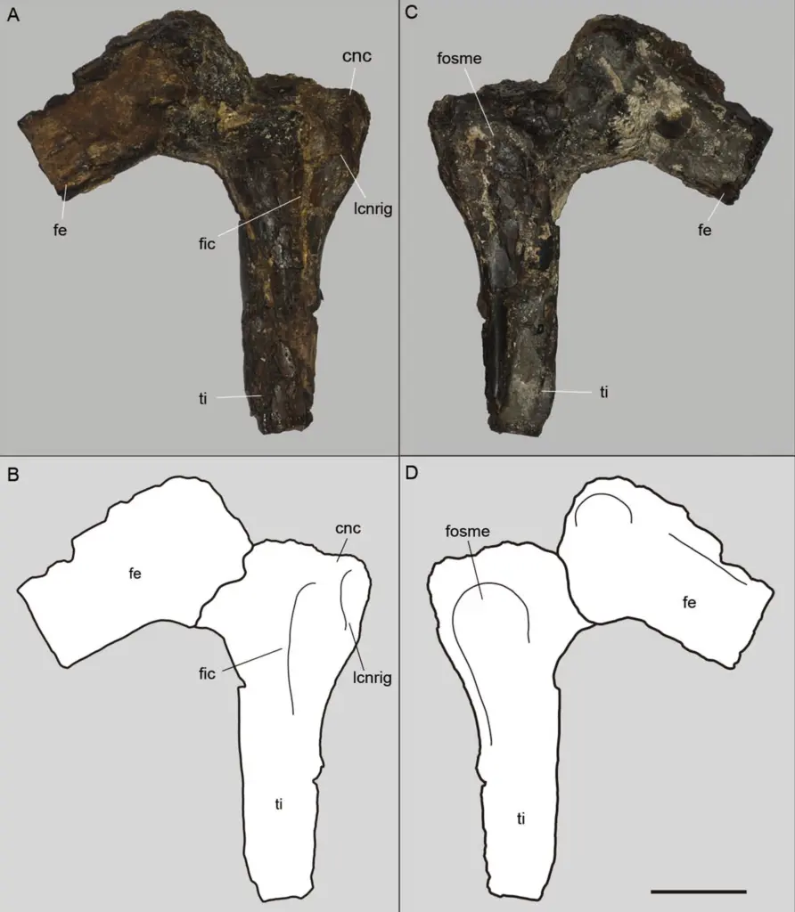
Kliti Grice and colleagues say they’ve found it: the oldest‑ever dinosaur red blood cells, from the hind limb of the Brazilian theropod Aratasaurus museunacionali, alongside chemical signatures of heme, the oxygen‑binding component of hemoglobin.
Who did this, and where it ran
The work comes from Kliti Grice at Curtin University and collaborators, and is published in the Proceedings of the National Academy of Sciences. In the study, the team reports red‑blood‑cell‑like microstructures and associated heme‑based chemical signals in limb bones from an animal about 10 feet (3 meters) long.
What they actually measured and how
This wasn’t a hunt for picturesque cell shapes alone. The researchers used chemical analysis to look for heme‑based signatures inside the fossil bone itself, tying chemistry to structures in place rather than to molecules extracted in bulk. A key tool was resonance Raman spectroscopy, which can amplify vibrational signals from specific ring‑shaped molecules like heme when illuminated at resonant wavelengths.
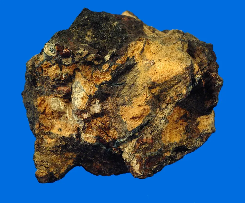
They also assessed the mineral context of the cellular look‑alikes. The team reports that the red‑blood‑cell‑like structures are mineralized with hausmannite, a manganese oxide (Mn₃O₄), and that the fossil preserves amino acids associated with collagen, the structural protein of bone.
Burial conditions that helped a blood trace hang on
Grice’s team argues the dinosaur’s rapid burial under sediment in a low‑oxygen setting, with microbial mats present, altered normal decay. Early mineralization could have stabilized or replicated microscopic biological structures before they were destroyed, while leaving altered but detectable chemical traces such as heme‑related compounds.
So, what did they find—numbers and names, not vibes
From limb bones of a roughly 10‑foot (3 meters) theropod dated to 115 million years ago, the team reports: (1) microscopic, red‑blood‑cell‑like structures; (2) in situ heme‑based chemical signatures consistent with blood components; (3) mineralization of those structures as hausmannite (Mn₃O₄); and (4) preserved amino acids associated with collagen. The authors describe this as the first study to use chemical processes to identify heme‑based signatures in dinosaur bones, and as the oldest chemical evidence of blood‑related material in a dinosaur fossil.
| Aspect | Details |
|---|---|
| Fossil age | 115 million years [c1] |
| Specimen | Hind‑limb of theropod Aratasaurus museunacionali [c2] |
| Animal size | ~10 feet (3 meters) long [c10] |
| Primary claim | Oldest‑ever dinosaur red blood cells [c2] |
| Key method | Resonance Raman spectroscopy; chemical identification of heme‑based signatures in bone [c3, c9] |
| Mineralization | Red‑blood‑cell‑like structures mineralized with hausmannite (Mn₃O₄) [c5] |
| Other biomolecules | Amino acids associated with collagen detected [c6] |
| Burial context | Rapid sediment cover, low oxygen, microbial mats [c4] |
Why not everyone is convinced yet
Mary Schweitzer, a molecular paleontologist not involved in the work, urges caution. She notes the structures do not convincingly match expected red blood cells in nonmammalian animals, which typically retain a nucleus that creates a visible bulge.
According to Schweitzer, the team’s images show more concave shapes with no nuclear bulge, and the structures are smaller than most non‑mammalian red blood cells. In her view, morphological similarity is not met—even if chemical signals are intriguing.
What this would mean if it holds up
If the heme signatures and cell‑like forms are truly endogenous, it pushes the record of dinosaur blood components back to 115 million years and strengthens the case that bone can preserve traces of original biochemistry. That, in turn, would widen the kinds of questions we can ask of fossils—about physiology, oxygen transport, and tissue organization—beyond what skeletal shape alone can tell us.
The deeper cut
How resonance Raman ‘listens’ for heme
Raman spectroscopy measures inelastic scattering of photons by molecular vibrations; resonance Raman further boosts bands from chromophores when the laser wavelength hits an electronic transition. Heme’s porphyrin ring is an ideal target: under resonance conditions, its core marker bands (e.g., C–C and C–N stretching in the macrocycle; out‑of‑plane modes tied to planar distortion) intensify by orders of magnitude relative to the background matrix. That selectivity is why researchers reach for resonance Raman when they suspect intact or modified porphyrins. The catch in fossils is spectral crowding and diagenetic overprints. Iron oxides and manganese oxides (here, hausmannite, Mn₃O₄) can fluoresce or contribute their own broad features, while geochemically altered porphyrins shift band positions and widths. Analysts lean on band pattern matching rather than single peaks, check for iron‑porphyrin signatures versus free porphyrins, and compare on‑ and off‑resonance spectra to discriminate genuine heme‑related signals from mineral or humic backgrounds. In situ measurements are critical: a spectrum co‑registered with a particular microstructure carries more weight than a bulk extract that can’t be tied to a cell‑sized feature.
What we still don’t know
Two big uncertainties remain. First, are the microstructures truly red blood cells from a nonmammalian dinosaur, given the lack of an obvious nuclear bulge and their small size? Second, how uniquely diagnostic are the reported heme‑based signals in this mineral context—could look‑alike spectra arise from diagenetic products or contamination? Answering both will likely require more specimens of Aratasaurus and other dinosaurs analyzed in situ with the same chemistry.
Why the preservation story matters
The team attributes the survival of these traces to rapid burial under low oxygen with microbial mats. Early mineralization—specifically, hausmannite coating or infilling—may have stabilized structures and shielded associated chemistry long enough for it to be partially preserved. If that recipe is right, it points paleontologists to similar depositional settings when prospecting for biochemical holdovers in deep time.
The paper: Heme signatures in mineralized red blood cells within vascular canals of a 115-million-year-old dinosaur bone (Proceedings of the National Academy of Sciences, 2026)
Related research: Blood to Molecules: The Fossil Record of Blood and Its Constituents (Topics in Geobiology, 2021)
Sources: Dinosaur red blood cells recovered from 115-million-year-old fossil (www.newscientist.com); Traces of ancient blood found in 115 million-year-old dinosaur fossil, new study claims (www.livescience.com)
Images: Cover: Everyman Science (illustration); Figure 1: Juliana Manso Sayão, Antônio Álamo Feitosa Saraiva, Arthur Souza Brum, Renan Alf / Wikimedia Commons (CC BY 4.0); Figure 2: Nessa Eull / Wikimedia Commons
How this article was made: Everyman Science uses AI tools to structure, format and optimise its articles, and occasionally to produce illustrations where no free photograph exists. The reporting these articles are based on is human-produced and cited above. Spotted an error? Write to [email protected] and we will correct it. — The editors How we work.





Science desk team of Everyman Science, curating and reporting on the day’s most significant developments in research, space exploration, and technology.